In a groundbreaking discovery, researchers in China have identified a previously unknown lymphatic-like system in the brain, upending conventional wisdom about brain physiology.
Historically, medical science believed that the neurovascular bundles that supply organs with nerves, arteries, veins, and lymphatic vessels operated independently of the brain.
Now new research has exposed the presence of the glymphatic system, a network that promotes the disposal of the brain’s waste products.
Decades in the Making
The long road that led to this revelation began as far back as the late 1960s when M. Földi and colleagues first described “pre-lymphatic channels” in the human brain, connecting to cervical lymph nodes.
It took another 40 years before researchers – Jeffrey Iliff and his colleagues – floated the idea of a glymphatic system in 2012. The researchers posited that this system managed the flow of cerebrospinal fluid through the brain, facilitating the removal of waste products, including synaptic vesicular debris and neuronal waste.
The research indicates that glymphatic flow, which accelerates while we sleep, plays a vital role in maintaining a healthy brain. When this flow is interrupted, waste will accumulate, potentially contributing to diseases such as Alzheimer’s, characterized by the accumulation of β-amyloid in the brain.
Measuring the Glymphatic Flow in the Brain
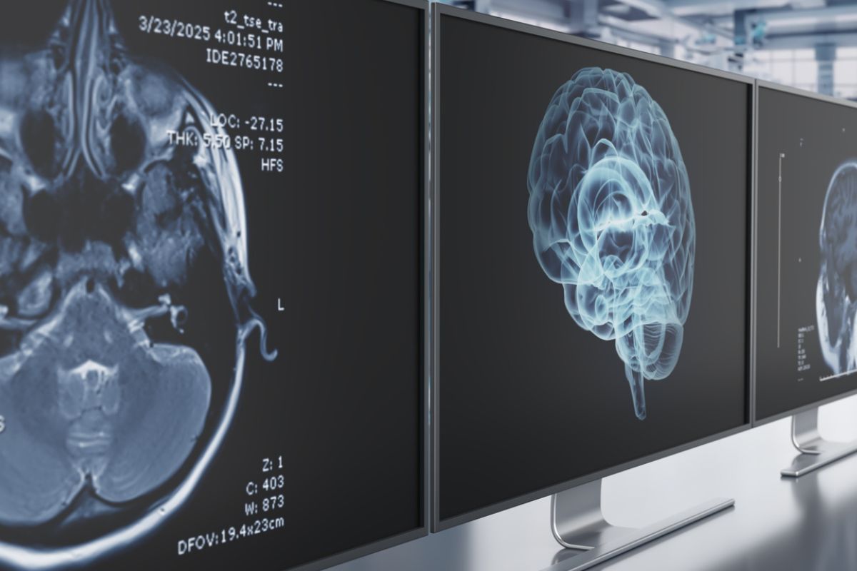
Despite the challenges of measuring glymphatic flow because of how slowly it works its way through brain tissue, researchers have developed an indirect approach that takes advantage of magnetic resonance imaging (MRI). These methods, including the calculation of perivascular space volume fraction (PVSVF-WM), free water (FW-WM) metrics, and the analysis along the perivascular space (ALPS) index, offer insights into the flow of the brain’s interstitial fluid.
In the latest issue of Radiology, Shiwei Lin and his colleagues focus on how obstructive sleep apnea (OSA) disrupts glymphatic flow. Researchers theorized that this disorder, which makes sleep difficult (at best), progressively affects glymphatic metrics with growing severity.
Methodology – And Hope
The study involved 105 participants with snoring issues and 50 healthy controls. Through extensive cognitive testing and MRI analysis, the researchers found a link between severe OSA and abnormal glymphatic metrics and impaired cognitive performance.
Later, a subset of participants with moderate OSA received a surgical procedure to address their symptoms. Post-surgery, these participants showed normalized glymphatic metrics and improved cognitive performance, hinting at the potential correction of glymphatic flow disruption.
While the researchers concede that further technological advancements would be required to measure glymphatic flow directly, the findings nevertheless open the door to early diagnosis and treatment of neurodegenerative diseases.
“The take-home message for radiologists is that this study provides strong evidence that relatively crude MRI measurements of glymphatic flow are probably good enough to use as biomarkers for diseases that accumulate waste products within the brain parenchyma,” John Port, MD, professor of radiology and associate professor of psychiatry at the Mayo Clinic in Rochester, Minnesota, wrote in a commentary about the research. “Alzheimer’s disease, Parkinson’s disease, multiple sclerosis, prion disease — we definitely need biomarkers to diagnose these diseases at an earlier stage so that we can begin treatment earlier and improve outcomes.”
Further Reading
Researchers Uncover Five Different Types of Alzheimer’s
Identifying and Treating Insomnia in Patients Living With Alzheimer’s