LESSONS LEARNED AT THE INTERFACE OF MEDICINE AND PSYCHIATRY
The Psychiatric Consultation Service at Massachusetts General Hospital sees medical and surgical inpatients with comorbid psychiatric symptoms and conditions. During their twice-weekly rounds, Dr Stern and other members of the Consultation Service discuss diagnosis and management of hospitalized patients with complex medical or surgical problems who also demonstrate psychiatric symptoms or conditions. These discussions have given rise to rounds reports that will prove useful for clinicians practicing at the interface of medicine and psychiatry.
Prim Care Companion CNS Disord 2022;24(6):22f03277
To cite: Donovan AL, Vyas CM, Petriceks A, et al. Agitation and an altered mental status in the emergency department: differential diagnosis, evaluation, and treatment. Prim Care Companion CNS Disord. 2022;24(6):22f03277.
To share: https://doi.org/10.4088/PCC.22f03277
© 2022 Physicians Postgraduate Press, Inc.
aDepartment of Psychiatry, Massachusetts General Hospital, Harvard Medical School, Boston, Massachusetts
bHarvard Medical School, Boston, Massachusetts
‡All authors contributed equally to the work.
*Corresponding author: Abigail L. Donovan, MD, 151 Merrimac St, Ste 501, Boston, MA 02114 ([email protected]).
Have you ever wondered why acute mental status changes develop and when they suggest an underlying neurologic disorder or drug-induced etiology? Have you been unsure about which aspects of the history, physical examination, and laboratory tests are most likely to yield meaningful information about etiology and treatment? Have you been uncertain about how (and why) you should manage acute agitation in the emergency department (ED)? If you have, the following case vignette and discussion should prove useful.
CASE VIGNETTE
Mr A, a disheveled 62-year-old man, was brought to the ED by police at the request of his wife because of his increasingly irritable and disturbing behavior over the past month. He described seeing little men walking through the walls and sitting in his living room; moreover, he believed that they were stealing his money and car keys. His medical history was notable for having type 2 diabetes mellitus, hypertension, and coronary artery disease. Since his wife thought that he was increasingly unreliable, she had been administering his medications to him over the past several months. Both he and his wife reported that he had never been evaluated by a psychiatrist.
When the ED physician attempted to perform a physical examination, Mr A screamed loudly, yelled obscenities, and made threatening gestures. Feeling unsafe, the physician called for the assistance of hospital security officers to ensure his own safety, that of Mr A, and others in the ED. Strategies to provide for the safety of all concerned became a priority; medications and restraints were ordered.
Mr A’s wife was interviewed in a separate area of the ED. She reported that her husband had been in his usual state of health until approximately 3 months earlier when he became more disorganized; he repeatedly lost his wallet and car keys and missed meetings at work. At times, he seemed inattentive and appeared somnolent. He developed clumsiness (bumping into furniture frequently and falling several times, although he had not hit his head or lost consciousness). Over the past month, he described seeing little men in their home who were stealing his wallet and keys. He had become progressively more irritable, and, on occasion, Mrs A worried that he might become violent. He had vivid dreams, and at times he acted them out while asleep. Mrs A confirmed that he was taking his medications, which included metformin, lisinopril, and atorvastatin; none of them had been changed recently. She reported that he drank 1 beer each week while watching football on television and that he did not use nicotine or recreational drugs. She mentioned that his father had developed odd behavior, hallucinations, a slow gait, and dementia in his 70s.
After Mr A was sedated, his blood was drawn for laboratory testing. His complete blood count (CBC) and differential, electrolytes, liver function tests (LFTs), thyroid function tests (TFTs), B12 level, folate level, rapid plasma regain (RPR) for syphilis, serum toxicology, urine toxicology, and urinalysis were all within normal limits. An electrocardiogram (EKG) and chest x-ray were also normal. His vital signs were monitored and were notable only for mild tachycardia, with a heart rate between 105 and 110 beats/minute. Several hours later, Mr A was calmer and able to participate with a physical examination. Notable findings included general bradykinesia and a bilateral pill-rolling tremor, but there were no other focal neurologic deficits. His mental status examination was notable for the absence of depression or mania; however, he described well-formed visual hallucinations. He denied having thoughts of suicide or homicide, and he had no insight into his symptoms. His Mini-Mental State Examination (MMSE)1 was 23 of 30, with points subtracted for “serial 7s,” following multistep directions, and copying a figure.
With this information, a differential diagnosis for his abnormal mental status and behavior was created, and further evaluation of his symptoms was pursued.
DISCUSSION
What Is Meant by Altered Mental Status?
Altered mental status (AMS) is not a diagnosis; instead, it is a clinical term that describes a wide range of signs and symptoms.2 Moreover, its meaning depends on the context in which it is used. AMS can present “as changes in consciousness, appearance, behavior, mood, affect, motor activity, or cognitive function.”3(p461)
One approach to understanding AMS is to organize the manifestations according to the components of the mental status examination.3 One might describe AMS by its behavioral signs (eg, abnormal appearance or behavior) that can be detected through observation.3 Patients may manifest poor hygiene; minimal attention paid to personal appearance, comfort, or safety; or behavior that is inappropriate for the context (eg, acting guarded and shouting at others when in a safe environment).4 AMS might reflect an altered mood or affect.4 Facial expressions can convey an elated mood, such as in a manic episode; mood may be fearful, such as when experiencing delusions of persecution; or affect may be flat, such as in schizophrenia. AMS could also refer to motoric manifestations, such as abnormal or unexpected gestures or postures or movements that are faster or slower than expected.4
Alternatively, AMS may involve impaired cognition, such as disorientation (ie, an inability of the patient to correctly state their name, location, or the current date).5 AMS may also indicate a deficit in attention (as demonstrated by an inability to count backward by 7s or to name the days of the week backward, among other tasks).5 AMS may refer to an alteration in language, whether in fluency, semantics, reading, writing, or other components.5 It may reflect a disturbance in memory, such as the inability to remember 3 words after 5 minutes or the inability to remember what one had for breakfast that morning. It may refer to impairments in executive function, planning, or visuospatial domains. In addition, it may suggest a change in thought content (as with hallucinations or delusions) or thought process.3 Since AMS is based on dysregulated affect, behavior, or cognition, clinicians need to create a broad differential diagnosis and be as clear as possible when describing its signs and symptoms.
What Can Cause Altered Mental Status?
Just as there are myriad meanings for AMS, there are a bevy of causes for the associated disparate presentations. Several clear and workable frameworks can assist clinicians to identify the causes of AMS.6
The differential diagnosis for AMS includes metabolic and medication-related etiologies (eg, excess use of opioids, benzodiazepines, steroids) and sequelae of other drugs (eg, alcohol, cocaine, marijuana).6 Changes in electrolytes may also precipitate AMS as a result of acute alterations (eg, hyponatremia in primary polydipsia) or underlying conditions (eg, hyponatremia secondary to malignancy). Therefore, clinicians should assess levels of sodium, potassium, calcium, and magnesium and pursue a workup as indicated. Endocrine derangements (eg, hyperthyroidism, diabetic ketoacidosis) may also lead to AMS. Environmental factors can impact metabolic activity by causing hypothermia, hyperthermia, or changes in oxygen saturation (eg, associated with high altitudes). Vitamin deficiencies (eg, pellagra, known by the “3 Ds” of diarrhea, dermatitis, and dementia, secondary to niacin deficiency or thiamine deficiency leading to Wernicke encephalopathy) can also precipitate AMS. Hepatic or uremic encephalopathy secondary to liver or renal dysfunction, respectively, can cause a wide constellation of findings associated with AMS.
Infections also induce AMS.6,7 Sepsis, meningitis, brain abscesses, and other systemic effects of infection (eg, urinary tract infection, pneumonia) can lead to changes in behavior and cognition. This may be of particular concern in the elderly, whose AMS may be mistaken for the cognitive impairment of dementia.8 Noninfectious inflammatory, autoimmune, and neurodegenerative conditions may also lead to AMS.6 Seizures of varying etiologies (eg, epilepsy, metabolic derangement, drug withdrawal) can cause AMS in the ictal or postictal state.
Primary neurologic and psychiatric conditions may cause AMS. Ischemic and hemorrhagic strokes are one important etiology, as are traumatic causes or brain injury.9 Tumors in the central nervous system (CNS) or the paraneoplastic effects of non-CNS tumors should also be considered.6 Finally, a wide range of primary psychiatric disorders such as mood disorders, psychotic disorders, various etiologies of dementia, and hyperactive or hypoactive catatonia should be included in the differential diagnosis. The main mood disorders to consider are bipolar type I and II and major depressive disorder.10 The main psychotic disorders to consider are schizophrenia, schizophreniform disorder, brief psychotic disorder, and delusional disorder.11 Dementias secondary to Alzheimer’s disease, vascular dementia, or neurodegenerative disorders such as Parkinson disease and diffuse Lewy body disease (DLBD) can also be associated with AMS.
Abnormalities in every organ system can lead to AMS; however, some of those etiologies are rapidly progressive and potentially lethal and should be considered first. Timely recognition and effective interventions will save lives.
Which Aspects of a Patient’s History (including medication use) Influence the Differential Diagnosis and Workup?
Agitation and an altered mental status can be triggered by a variety of underlying causes, including substance intoxication or withdrawal (from illicit or prescribed medications), psychiatric illness, and medical illness.12 Several aspects of the presentation, including the timeline, the underlying psychiatric and medical history, the associated symptoms, and the family history can further refine the differential diagnosis. Patients may also have more than 1 illness that contributes to their mental status changes; therefore, it is important to maintain a broad differential.13 If a patient is unable to provide historical information or if the accuracy of information is suspect, collateral information from family, friends, or health care providers will be critical. Collateral information from family members should be gathered away from the patient so that they can speak freely.
Understanding the timeline and course of the clinical presentation is crucial. Symptoms with an acute onset (ie, over hours to days) suggest an acute medical event (such as a cerebrovascular accident, substance or medication intoxication or withdrawal), while those with an acute onset with a fluctuating course are suggestive of delirium. A subacute course (ie, over days to weeks) suggests a condition that develops gradually (like mania or a psychotic decompensation in schizophrenia). A more chronic course (ie, persisting over several months) is suggestive of a longer disease process such as dementia (including Alzheimer’s disease, DLBD, and frontotemporal dementia). A stepwise and chronic course suggests a vascular dementia.14 Finally, an acute on chronic course (several months or years of impairment, with an acute worsening over hours to days) suggests that 2 or more illnesses may be present, as is the case with a delirium superimposed on dementia.
Multiple aspects of a patient’s history can help to inform this differential diagnosis. The patient’s medical and psychiatric history may provide important clues. A history of medical illnesses that can be associated with acute mental status changes (such as endocrinopathies, pulmonary disease with hypoxia, and severe hypertension) should raise one’s suspicion for delirium. A history of psychiatric illnesses associated with acute mental status changes (such as schizophrenia, bipolar disorder, or dementia) should prompt consideration of a psychiatric etiology. Similarly, a history of a substance use disorder should raise suspicion for intoxication or withdrawal states. Prescribed medications must also be reviewed. Many medications have a narrow therapeutic window, wherein small changes in dosages or blood concentrations can lead to significant adverse effects, leaving patients vulnerable to toxicity. Overdose and abrupt discontinuation can both be associated with agitation and acute mental status changes. The age of the patient is also important. As patients age, their risk of developing a new mood or psychotic disorder decreases, while their risk of developing dementia increases.
A detailed understanding of the co-occurring symptoms can also help narrow the differential diagnosis. For example, agitation that occurs in the context of a waxing and waning mental status, with fluctuations over the course of the day, as well as inattention and impaired consciousness, is highly suggestive of delirium. Alternatively, agitation that occurs in the context of a decreased need for sleep, pressured speech, hypersexuality, impulsivity, and euphoria is suggestive of bipolar disorder. Comorbid symptoms also suggest a toxidrome; for example, mydriasis, dry skin, urinary retention, hyperthermia, tachycardia, and hypertension suggest anticholinergic toxicity. Agitation occurring in the context of memory changes or declining executive function should raise consideration of a dementing illness.
In addition, a social and occupational history should be obtained to assess premorbid intelligence, cognitive function, and level of education (which influences subsequent mental status examinations) and to screen for environmental risk factors. Finally, family history can indicate a patient’s vulnerability to developing an illness or group of illnesses, including dementias, mood disorders, and schizophrenia.
In the case of Mr A, the most notable findings included a subacute onset over several months (rather than hours to days), making delirium unlikely. The lack of a psychiatric and family psychiatric history make a mood disorder or psychotic disorder less likely. His medications were unlikely contributors, as was his limited use of alcohol. However, his age, family history, and the time course all elevate dementia within the differential diagnosis.
Which Aspects of the Physical Examination Should Be Assessed in Those With Altered Mental Status?
The physical examination of a patient with AMS must be systematic and thorough. Even in cases in which a psychiatric cause is suspected, a thorough physical examination is warranted to exclude potential contributing or causative factors.
Complete vital signs (including heart rate, blood pressure, respiratory rate, and oxygen saturation) are crucial, as specific alterations can help to identify an underlying illness. The physical examination should be completed with special attention paid to associated signs or symptoms that might indicate a diagnosis. For example, the presence of Argyll-Robinson pupils indicates neurosyphilis, while the co-occurrence of ataxia and ophthalmoplegia strongly suggests Wernicke-Korsakoff syndrome. Findings of the physical examination can also indicate a toxidrome (eg, dry skin and mydriasis are seen in anticholinergic toxicity, which would be relevant in the case of an acute mental status change that develops over several hours).15
A neurologic examination is also critical. The complete examination should include an assessment of cranial nerves, sensory and motor function, deep tendon reflexes, cerebellar function, and gait.16 Focal findings suggest specific etiologies, such as an acute stroke or Parkinson disease. Screening for visual or hearing impairment should be conducted, as these sensory deficits can exacerbate cognitive and behavioral changes.16 In the case of Mr A, his physical examination was most notable for the presence of parkinsonian symptoms (including bradykinesia and tremor), without other focal deficits suggestive of other neurologic illnesses.
Which Laboratory Tests Should Be Conducted in the Emergency Department When a Person Presents With Agitation and Altered Mental Status?
Laboratory testing must be broad in scope and include assessment of reversible causes of mental status changes, including vascular (stroke), infectious (neurosyphilis), neoplastic (primary, metastatic, paraneoplastic), degenerative (multiple sclerosis), inflammatory (vasculitis), endocrine (hyperthyroidism or hypothyroidism), metabolic (thiamine deficiency), toxic (medication effects), traumatic (dementia pugilistica), and other (normal pressure hydrocephalus) etiologies.16 At a minimum, laboratory testing should include a CBC with a differential, a chemistry panel (with calcium, magnesium, phosphorus, and glucose), LFTs, TFTs, and levels of B12 and folate, as well as an RPR, applicable serum drug levels (eg, of digoxin, lithium, alcohol), a urine drug screen, and a urinalysis.17 An EKG and chest x-ray, a computed tomography (CT) scan, or a magnetic resonance imaging (MRI) scan should be obtained. An MRI is preferred unless an intracranial hemorrhage is suspected. Additional testing is needed if the initial workup is unrevealing or if atypical symptoms are present. For example, a lumbar puncture should be performed to obtain cerebrospinal fluid for analysis if there are concerns for CNS infection or autoimmune encephalitis. An electroencephalogram (EEG) can help to diagnose a seizure disorder or Creutzfeldt-Jacob disease or to confirm a diagnosis of delirium.16 In the case of Mr A, since the initial laboratory investigation revealed no abnormalities, further diagnostic testing was conducted.
What Are the Life-Threatening Causes of Altered Mental Status?
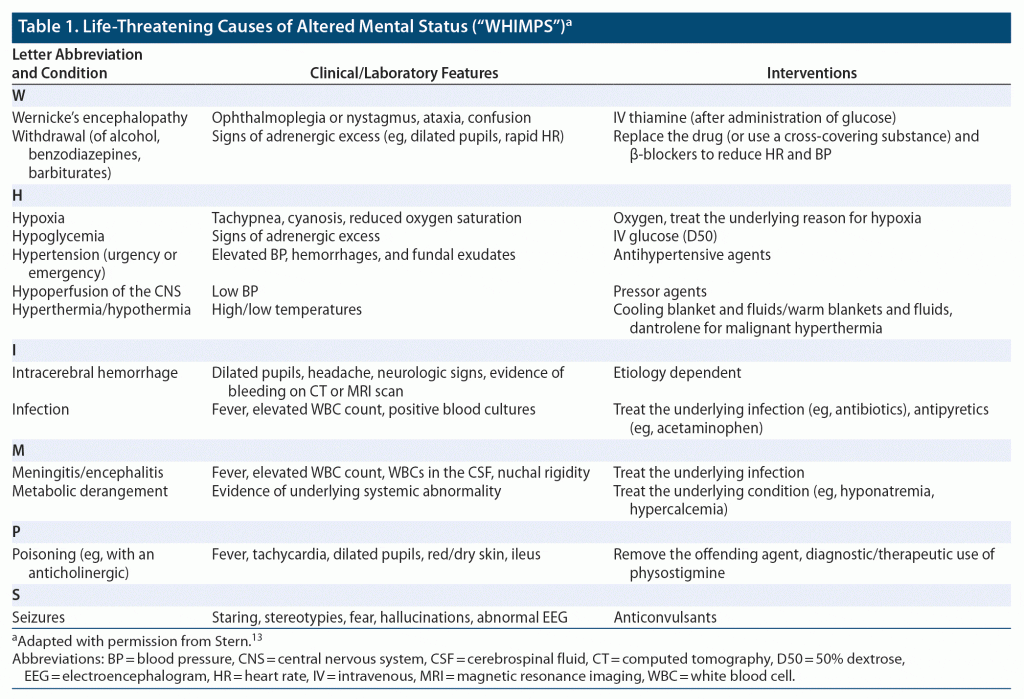
Although any illness (localized or systemic) or physiologic stress can contribute to AMS (with disturbances in affect, behavior, and cognition), in general, the greater the disturbance caused by the process, the greater the risk of altered CNS function. While the primary disturbance can involve 1 or more organ systems (eg, CNS, cardiopulmonary, endocrine, metabolic, rheumatologic/musculoskeletal, hematologic, gastrointestinal, urogenital), some conditions can be lethal if left undiagnosed and untreated in a timely fashion. These conditions can be recalled by the mnemonic, “rule out the WHIMPS,” with each letter of the mnemonic representing a different condition (Table 1).18 Fortunately, each of these conditions can be assessed quickly, which can lead to life-saving interventions.
Which Conditions Are More Often Associated With Visual Hallucinations?
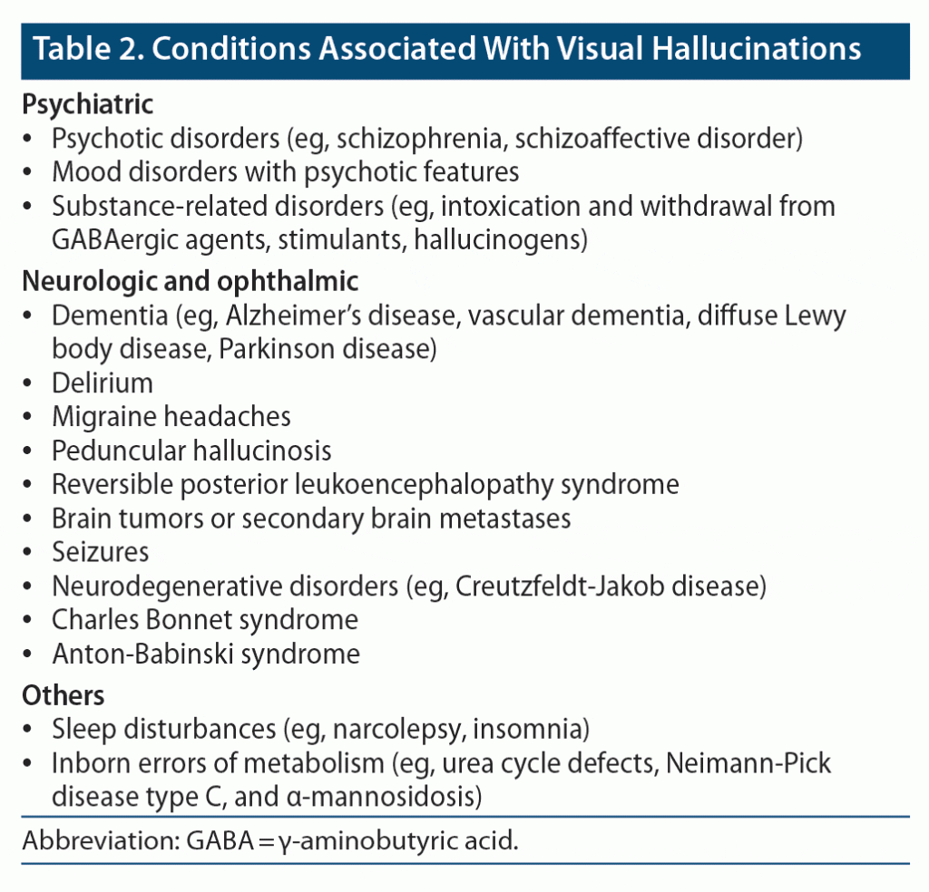
Hallucinations, which can occur with any of the 5 senses (hearing, seeing, smelling, tasting, feeling), are perceptions of nonexistent events or objects in the absence of an external stimulus. They can arise with dysfunction of any organ system (eg, CNS, cardiopulmonary, endocrine, hematologic), as well as psychiatric, medical, metabolic, and ophthalmic disorders, and with use of or withdrawal from a variety of drugs (Table 2).19 Although auditory hallucinations have been described most frequently in individuals with primary psychiatric disorders (eg, schizophrenia), other forms of hallucinations are common. Conditions with a neurologic etiology are more apt to be linked with visual hallucinations than are hallucinations related to the other senses. Among individuals with migraines, approximately one-third experience visual hallucinations before their headache develops (the “aura”).20 In addition, clinical and translational research in ophthalmology has determined that visual hallucinations are one of the hallmarks of Charles Bonnet syndrome—a condition that arises from damage to the visual system (eg, age-related macular degeneration) or deafferentation of the visual cortex (interruption of the nerves that carry input to the visual cortex).21,22 Patients with sleep disturbances (eg, insomnia, narcolepsy) are more likely to experience visual hallucinations at sleep onset (hypnagogic hallucinations) or upon waking (hypnopompic hallucinations).23
Symptoms of psychosis have been reported in 15%–78% of people with major neurocognitive disorders,24 often correlated with the etiology of dementia. Notably, early onset visual hallucinations occur in as many as 80% of patients with DLBD; thus, the presence of visual hallucinations helps to differentiate DLBD from Parkinson disease and Alzheimer’s disease (in which visual hallucinations are less prevalent).
Who Develops Paranoia?
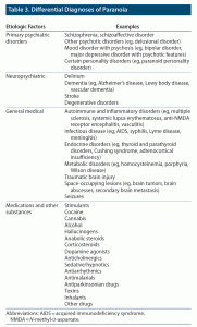
Paranoia, a symptom comprised of extreme suspiciousness and mistrust of others, can develop into delusions (ie, fixed, false beliefs) of persecution, jealousy, or imminent harm. On a population level, paranoia is found across the spectrum of severity. For example, mild paranoia is relatively common among individuals under intense stress. However, paranoia can evolve into irrational, uncontrollable, or long-lasting thoughts and lead to hazardous actions (such as physical aggression). When this type of paranoia develops, it should become a focus in clinical care, and protection of the patient experiencing the paranoia and others may be required. Paranoia arises from a confluence of risk factors, including biological conditions (eg, medical and neurologic disorders, adverse reactions to medications, family history), sociocultural features (eg, race/ethnicity, social support), environmental factors (eg, migration, toxicologic), and clinical conditions (eg, primary psychiatric etiologies).25,26 Table 3 illustrates differential diagnoses of paranoia. Among the conditions listed in Table 3, severe and persistent paranoia is a central feature of primary psychiatric disorders, including psychotic and mood disorders. A comprehensive assessment with a detailed psychiatric evaluation is needed to determine the underlying etiology of paranoia. For example, paranoid delusions often coexist with other psychotic symptoms (eg, disorganized speech, hallucinations) in individuals with schizophrenia. Conversely, those with a persecutory type of delusional disorder may not have other psychotic symptoms or impairments in psychological functioning. In addition, a genetic predisposition (eg, family history) to paranoia and childhood abuse or neglect are associated with an elevated risk of developing a paranoid personality disorder27; early recognition of these risk factors may guide prevention and intervention strategies.
Psychotic symptoms can arise in diverse neuropsychiatric and medical conditions (Table 3).28,29 Paranoia due to a nonpsychiatric etiology may exhibit atypical features (eg, fluctuations in cognitive performance), as well as abnormal vital signs or laboratory tests. Illicit drug use is the most common cause of secondary paranoia, especially in adolescents and young adults; in addition, new-onset paranoia can be due to drug withdrawal.30 Elderly individuals are at elevated risk for secondary psychosis (and paranoia) due to drugs, toxins, use of polypharmacy, and underlying medical illnesses.
Which Treatments Can Reduce Hallucinations and Paranoia?
Although treatment of hallucinations and paranoia is predicated on its etiology, symptomatic treatments may be required before the etiology has been established. Immediate safety measures might be necessary for someone with a command auditory hallucination to hurt themselves or others or someone with intense persecutory delusions presenting as agitated. In those situations, immediate measures are necessary to ensure the safety of the patient and others. In addition to implementing safety protocols, physical or chemical restraints might be needed. Parenteral haloperidol (with or without lorazepam), intramuscular (IM) olanzapine,31 and parenteral droperidol are all reasonable options.32 It is important to note that in 2005, the US Food and Drug Administration (FDA) issued an advisory warning on the concomitant administration of intramuscular olanzapine along with benzodiazepines due to the potential for excessive sedation and cardiorespiratory depression following publication of multiple postmarketing adverse events, including 29 fatalities, associated with IM olanzapine in the setting of IM benzodiazepines use.33,34 Yet, data supporting a strong clear cause and effect between olanzapine/benzodiazepine combinations have been inconsistent. Despite the FDA warning, olanzapine IM and lorazepam IM coadministration has been reported, with a 2018 study of 91 patients showing it to be safe without serious adverse events (eg, instances of hypotension, bradycardia, bradypnea, or oxygen desaturation) following administration.35
Once safety has been assured, either psychopharmacologic or neurotherapeutic agents can be employed to manage hallucinations or paranoia.36 A second-generation antipsychotic (SGA) medication (eg, olanzapine, risperidone, quetiapine) or a first-generation antipsychotic (FGA) (eg, haloperidol, fluphenazine) can be used to control psychotic symptoms. While evidence for the superiority of 1 agent over others to control agitation, hallucinations, or paranoia is lacking,37 the use of SGAs is increasingly recommended because of greater tolerability.38 The availability of orally dissolvable formulations of certain antipsychotics (like risperidone and olanzapine) allows for more assurance that the medication has been ingested. The agent used to control the symptoms depends on the treatment history of a patient, the clinician’s preference, or the hospital’s protocol. Depending on the severity of the symptoms and the response to the antipsychotic medication, augmentation with a benzodiazepine (eg, lorazepam) or a mood stabilizer (eg, sodium valproate) might be necessary to achieve patient stabilization.39,40 If a patient is uncooperative or a faster onset is desired, use of a parenteral antipsychotic or benzodiazepine should be considered. Clozapine is the drug of choice when hallucinations and paranoia are treatment resistant; such individuals will require further evaluation and close monitoring.41 Neurotherapeutic agents, especially electroconvulsive therapy, can be a very efficacious tool to control acute manifestations of hallucinations and paranoia in the ED.42
Which Adverse Side Effects Should Be Considered Before Antipsychotics Are Administered?
While use of antipsychotics may be required to rapidly manage acute agitation and aggressive behavior that can be life threatening and not responding to nonpharmacologic interventions, clinicians should be mindful of their possible side effects and how to manage them.32 Safety and efficacy of antipsychotics varies according to a patient’s individual characteristics (eg, age, medical history, pharmacologic history) and their acute presentation (eg, alcohol intoxication) as well as the antipsychotic agent used (eg, as the mechanism of action for them differs), the dosage used (1 time vs multiple doses over more days), and the route of administration (eg, oral, intramuscular, intravenous).8,32,43 Antipsychotic medications all achieve rapid sedation or tranquilization; however, they have significant differences in their side effects profile and prevalence.32 FGAs, such as haloperidol and droperidol, and SGAs, such as olanzapine, are the most frequently used antipsychotics for treatment of agitation in the ED.32
Some of the most common short-term side effects from administration of antipsychotics include excessive sedation, somnolence, hypotension, and dizziness, which increase the risk of falls (and that may lead to injuries and fractures), especially in the elderly.8,43 Of note, ziprasidone has been associated with the highest incidence of somnolence (20%) and nausea (12%).32 Acute involuntary movement disorders, such as akathisia and acute dystonia, are common and primarily related to use of FGAs (eg, haloperidol, droperidol). Coadministration of FGAs and benzodiazepines (eg, midazolam, lorazepam), or with benztropine or diphenhydramine, can lower the risk of acute extrapyramidal symptoms (EPS). However, EPS (eg, parkinsonism) can develop with use of SGAs (eg, olanzapine), especially after repeated doses over several days.44 QT interval prolongation is frequent with use of antipsychotics, especially with ziprasidone and haloperidol, but quite rare with olanzapine. While QT prolongation increases with repeated doses, it rarely leads to a corrected QT (QTc) > 450 msec. Torsades de pointes is rare and primarily associated with use of intravenous (IV) haloperidol.32 Both QT prolongation and Torsades de pointes are more likely to occur in patients with preexisting cardiac rhythm abnormalities and with use of food or drugs that affect the P450 isoenzyme system and delay metabolism of antipsychotics.
Airway compromise or other respiratory complications (eg, aspiration pneumonia, respiratory depression, hypoxia) associated with use of antipsychotics rarely require intubation; however, it is more likely to happen when SGAs are administered in patients with alcohol intoxication. For this reason, the FDA recommends against coadministration of antipsychotics and benzodiazepines to reduce the higher risk of excessive sedation and cardiopulmonary depression.32,35,45 Further, since ziprasidone may have teratogenic effects, its use should be avoided in women of childbearing age.46
The potential for side effects highlights the need for a thoughtful approach (based on patient presentation and the urgency for intervention) to the choice of antipsychotic medication. Often, a patient’s medical history might be unavailable, and the examination may be limited due to patient’s agitation and inability to engage safely in one. In these cases, ED clinicians should base their antipsychotic decision on the desired result, while continuously monitoring the patient for possible side effects so that timely interventions can be instituted should they become necessary. As a general rule, the lowest effective dose of an antipsychotic should be used to achieve clinical efficacy and safe sedation for the shortest time, rather than anticipating the need for long-term treatment; this strategy will reduce the frequency and severity of adverse side effects.8,43
Which Treatment Settings Can Be Used After Discharge From the Emergency Department?
Disposition of patients who present with AMS to the ED is as important as the acute management while in the ED. Deciding on the most appropriate level of care for the patient with AMS as they transition out of the ED depends on many factors, including the severity of the clinical presentation, its etiology, and how easily it can be identified, addressed, and reversed. In addition, it is crucial to know where the patient is compared to their baseline levels, the likelihood of this situation recurring soon, and the availability of adequate aftercare planning, support, and medical knowledge to safely manage the patient. A recent study47 found that patients with AMS who presented to the ED had a significantly higher risk of death within 7 days of their arrival than did patients who presented with chest pain and a significantly higher risk of dying within 30 days compared to patients with chest pain and headache. Of note, most patients with AMS require an inpatient hospitalization (eg, to monitor and manage their hemodynamics, airway, incapacity, and overall medical and psychiatric problems).8,48 Given the high mortality risk associated with AMS, the affective, behavioral, and cognitive manifestations of AMS should always be taken seriously and the etiology investigated. Moreover, the threshold for hospitalization for those with AMS should be low.
Patients with AMS in the ED often become easily frustrated, especially if they are boarding in the ED for a prolonged time (hours to days) while awaiting an inpatient bed; thus, they may ask to leave the ED against medical advice. This can present a safety risk for the patient with AMS and put ED physicians in the difficult position of wanting to respect the patient’s autonomy while questioning their capacity to make medical decisions. While rigorous capacity determination is important and necessary, so is the involvement of the patient’s family, friends, or next of kin who can advocate for the patient to remain in the hospital and receive much-needed care. A shared decision-making approach that includes empathically listening to the patient and his/her reasons for wanting to leave, while addressing what is possible regarding competing needs (eg, pet care, childcare, work) is important. Throughout this process, patients with AMS should be informed about the process they are facing and the time that might be needed; this can help patients stay in care and receive disposition recommendations.49
However, not all patients with AMS require hospitalization; some can be safely discharged to their home or to their place of primary residence. This disposition can be considered for those in whom AMS has resolved, and the underlying cause has been identified and reversed. Safe discharge can also be arranged when there are reliable caregivers and accessible transportation, and the patient is able to follow aftercare treatment plan recommendations. No matter which disposition setting has been arranged for the patient with AMS, timely and effective communication among the ED staff and the inpatient or outpatient provider will be important to coordinate care and ensure the patient’s safety.8
What Happened to Mr A?
Mr A was admitted to the neurology inpatient service for further evaluation of his mental status changes. He had an EEG that revealed decreased reactivity of background activity as well as frontal intermittent rhythmic delta activity. His MRI revealed mild diffuse frontal and parieto-temporal volume loss, pallor of the substantia nigra, and preserved mesial temporal lobe and hippocampal volume. He was diagnosed with DLBD and was started on rivastigmine and low-dose quetiapine, which reduced his agitation. One week later, he was discharged to his home with his wife, with close follow-up from the psychiatry and neurology departments.
How Does Diffuse Lewy Body Disease Usually Present and What Is Its Course?
Diffuse Lewy body disease, or dementia with Lewy bodies, is one of the most common causes of dementia among the elderly. Its main pathophysiologic features are proteinaceous deposits, called Lewy bodies, which are largely found in the cortex, amygdala, and brain stem. Clinical manifestations of the disorder are similar to those of Alzheimer’s disease; however, visual hallucinations, a fluctuating course, a sensitivity to antipsychotic medications, executive dysfunction, apathy, irritability, visuospatial and memory impairment, sleep disturbances (especially rapid eye movement behavior disorder), and agitation are common. Unfortunately, there are no FDA-approved treatments for the condition, and the average course (with substantial variability) of the disease is 5–7 years.50
CONCLUSION
Alterations in mental status (that reveal themselves in disturbances of affect, behavior, and cognition) are both prevalent and problematic. They can herald life-threatening emergencies and be attributable to dysfunction within every organ system of the body and can be viewed as medical, neurologic, or psychiatric. A diligent and comprehensive search for the cause of AMS is of utmost importance, as treatment is largely predicated on its etiology. While the search for the underlying problem is being conducted, symptomatic interventions may be necessary to ensure the safety of the patient, family members, and medical staff. Being mindful of the side effects of pharmacologic treatments can help to guide the choice of agents used, as can the etiology of AMS, its severity, the response to medications (eg, antipsychotics, benzodiazepines, mood stabilizers), and the routes of administration available.
Submitted: March 3, 2021; accepted April 29, 2022.
Published online: December 8, 2022.
Relevant financial relationships: Dr Vyas has received research salary support from Nestlé Purina PetCare Company. Drs Donovan, Paudel, Van Alphen, and Stern and Mr Petriceks report no relevant financial relationships.
Funding/support: None.
Clinical Points
- Altered mental status (AMS) is not a diagnosis; instead, it is a clinical term that describes a wide range of signs and symptoms.
- Abnormalities in every organ system can lead to AMS and can be considered medical, neurologic, or psychiatric.
- Choice of treatment for AMS depends on its etiology, severity, response to medications (eg, antipsychotics, benzodiazepines, mood stabilizers), and available routes of administration.
- Timely and effective communication among the emergency department staff and the inpatient or outpatient provider are important to coordinate care and ensure the patient’s safety.
References (50)

- Folstein MF, Folstein SE, McHugh PR. “Mini-mental state”: a practical method for grading the cognitive state of patients for the clinician. J Psychiatr Res. 1975;12(3):189–198. PubMed CrossRef
- Xiao HY, Wang YX, Xu TD, et al. Evaluation and treatment of altered mental status patients in the emergency department: life in the fast lane. World J Emerg Med. 2012;3(4):270–277. PubMed CrossRef
- Veauthier B, Hornecker JR, Thrasher T. Recent-onset altered mental status: evaluation and management. Am Fam Physician. 2021;104(5):461–470. PubMed
- Norris D, Clark MS, Shipley S. The mental status examination. Am Fam Physician. 2016;94(8):635–641. PubMed
- Kipps CM, Hodges JR. Cognitive assessment for clinicians. J Neurol Neurosurg Psychiatry. 2005;76(suppl 1):i22–i30. PubMed CrossRef
- Burke J. Altered Mental Status. In: Saint S, Chopra V, eds. The Saint-Chopra Guide to Inpatient Medicine. Oxford University Press; 2018:463–466.
- Patti L, Gupta M. Change in Mental Status. In: StatPearls [Internet]. Treasure Island, FL: Stat Pearls Publishing; 2022.
- Han JH, Wilber ST. Altered mental status in older patients in the emergency department. Clin Geriatr Med. 2013;29(1):101–136. PubMed CrossRef
- Wilber ST, Ondrejka JE. Altered mental status and delirium. Emerg Med Clin North Am. 2016;34(3):649–665. PubMed CrossRef
- Sekhon S, Gupta V. Mood Disorder. In: StatPearls [Internet]. Treasure Island, FL: Stat Pearls Publishing; 2022.
- Lieberman JA, First MB. Psychotic disorders. N Engl J Med. 2018;379(3):270–280. PubMed CrossRef
- Tesar GE, Stern TA. The evaluation and treatment of agitation in the intensive care unit. J Intensive Care Med. 1986;1(3):137–148. CrossRef
- Stern TA. Learning About Agitation, Confusion, and Altered Mental Status: A Programmed Text for Multidisciplinary Learners. Boston, MA: Massachusetts General Hospital Psychiatry Academy; 2020.
- Chemali ZN, Stern TA. Facing Memory Loss and Dementia: A Guide for Patients and Their Families. Boston, MA: Massachusetts General Hospital Psychiatry Academy; 2021.
- Keary CJ, Nejad SH, Rasimas JJ, et al. Intoxications associated with agitation, tachycardia, hypertension, and fever: differential diagnosis, evaluation, and management. Prim Care Companion CNS Disord. 2013;15(3):PCC.12f01459. PubMed CrossRef
- Gatchel JR, Wright CI, Falk WE, et al. Dementia. In: Stern TA, Fava M, Wilens TE, et al, eds. Massachusetts General Hospital Comprehensive Clinical Psychiatry. 2nd ed. Philadelphia, PA: Elsevier; 2016.
- King F, Wiechers ER. Patients With Neurocognitive Disorders. In: Stern TA, Freudenreich O, Smith FA, et al, eds. Massachusetts General Hospital Handbook of General Hospital Psychiatry. 7th ed. Edinburgh: Elsevier; 2017
- Caplan JP, Cassem NH, Murray GB, et al. Delirium. In: Stern TA, Rosenbaum JF, Fava M, et al, eds. Massachusetts General Hospital Comprehensive Clinical Psychiatry. Philadelphia, PA: Mosby/Elsevier; 2008:217–229.
- Teeple RC, Caplan JP, Stern TA. Visual hallucinations: differential diagnosis and treatment. Prim Care Companion J Clin Psychiatry. 2009;11(1):26–32. PubMed CrossRef
- Launer LJ, Terwindt GM, Ferrari MD. The prevalence and characteristics of migraine in a population-based cohort: the GEM study. Neurology. 1999;53(3):537–542. PubMed CrossRef
- Pang L. Hallucinations experienced by visually impaired: Charles Bonnet syndrome. Optom Vis Sci. 2016;93(12):1466–1478. PubMed CrossRef
- Rovner BW. The Charles Bonnet syndrome: a review of recent research. Curr Opin Ophthalmol. 2006;17(3):275–277. PubMed CrossRef
- Ohayon MM. Prevalence of hallucinations and their pathological associations in the general population. Psychiatry Res. 2000;97(2-3):153–164. PubMed CrossRef
- Tampi RR, Young J, Hoq R, et al. Psychotic disorders in late life: a narrative review. Ther Adv Psychopharmacol. 2019;9:2045125319882798. PubMed CrossRef
- Dean K, Murray RM. Environmental risk factors for psychosis. Dialogues Clin Neurosci. 2005;7(1):69–80. PubMed CrossRef
- Freeman D, McManus S, Brugha T, et al. Concomitants of paranoia in the general population. Psychol Med. 2011;41(5):923–936. PubMed CrossRef
- Johnson JG, Cohen P, Brown J, et al. Childhood maltreatment increases risk for personality disorders during early adulthood. Arch Gen Psychiatry. 1999;56(7):600–606. PubMed CrossRef
- Keshavan MS, Kaneko Y. Secondary psychoses: an update. World Psychiatry. 2013;12(1):4–15. PubMed CrossRef
- Friedman JH. Parkinson disease psychosis: update. Behav Neurol. 2013;27(4):469–477. PubMed CrossRef
- Griswold KS, Del Regno PA, Berger RC. Recognition and differential diagnosis of psychosis in primary care. Am Fam Physician. 2015;91(12):856–863. PubMed
- Battaglia J. Pharmacological management of acute agitation. Drugs. 2005;65(9):1207–1222. PubMed CrossRef
- Kim HK, Leonard JB, Corwell BN, et al. Safety and efficacy of pharmacologic agents used for rapid tranquilization of emergency department patients with acute agitation or excited delirium. Expert Opin Drug Saf. 2021;20(2):123–138. PubMed CrossRef
- Zacher JL, Roche-Desilets J. Hypotension secondary to the combination of intramuscular olanzapine and intramuscular lorazepam. J Clin Psychiatry. 2005;66(12):1614–1615. PubMed CrossRef
- Marder SR, Sorsaburu S, Dunayevich E, et al. Case reports of postmarketing adverse event experiences with olanzapine intramuscular treatment in patients with agitation. J Clin Psychiatry. 2010;71(4):433–441. PubMed CrossRef
- Williams AM. Coadministration of intramuscular olanzapine and benzodiazepines in agitated patients with mental illness. Ment Health Clin. 2018;8(5):208–213. PubMed CrossRef
- Paudel S, Vyas CM, Peay C, et al. New-onset paranoia in an elderly woman with bipolar disorder: differential diagnosis, evaluation, and treatment. Prim Care Companion CNS Disord. 2022;24(4):21f03141. PubMed CrossRef
- Muir-Cochrane E, Oster C, Gerace A, et al. The effectiveness of chemical restraint in managing acute agitation and aggression: a systematic review of randomized controlled trials. Int J Ment Health Nurs. 2020;29(2):110–126. PubMed CrossRef
- Correll CU, Yu X, Xiang Y, et al. Biological treatment of acute agitation or aggression with schizophrenia or bipolar disorder in the inpatient setting. Ann Clin Psychiatry. 2017;29(2):92–107. PubMed
- Hovens JE, Dries PJ, Melman CT, et al. Oral risperidone with lorazepam versus oral zuclopenthixol with lorazepam in the treatment of acute psychosis in emergency psychiatry: a prospective, comparative, open-label study. J Psychopharmacol. 2005;19(1):51–57. PubMed CrossRef
- Tseng PT, Chen YW, Chung W, et al. Significant effect of valproate augmentation therapy in patients with schizophrenia: a meta-analysis study. Medicine (Baltimore). 2016;95(4):e2475. PubMed CrossRef
- Van Alphen MU, Vyas CM, Paudel S, et al. Managing clozapine in primary care settings. Prim Care Companion CNS Disord. 2022;24(1):21f02948. PubMed CrossRef
- Grover S, Sahoo S, Rabha A, et al. ECT in schizophrenia: a review of the evidence. Acta Neuropsychiatr. 2019;31(3):115–127. PubMed CrossRef
- Nikooie R, Neufeld KJ, Oh ES, et al. Antipsychotics for treating delirium in hospitalized adults: a systematic review. Ann Intern Med. 2019;171(7):485–495. PubMed CrossRef
- Chandrasena R, Dvoráková D, Lee SI, et al. Intramuscular olanzapine vs intramuscular short-acting antipsychotics: safety, tolerability and the switch to oral antipsychotic medication in patients with schizophrenia or acute mania. Int J Clin Pract. 2009;63(8):1249–1258. PubMed CrossRef
- Herzig SJ, LaSalvia MT, Naidus E, et al. Antipsychotics and the risk of aspiration pneumonia in individuals hospitalized for nonpsychiatric conditions: A cohort study. J Am Geriatr Soc. 2017;65(12):2580–2586. PubMed CrossRef
- Connor TH, MacKenzie BA, DeBord DG, et al. NIOSH list of antineoplastic and other hazardous drugs in healthcare settings. US Department of Health and Human Services, Centers for Disease Control and Prevention, National Institute for Occupational Safety and Health, DHHS (NIOSH). Publication number 2016–161. CDC/NIOSH website. Accessed November 3, 2022. https://www.cdc.gov/niosh/docs/2016-161/pdfs/2016-161.pdf
- Stanich JA, Oliveira J e Silva L, Ginsburg AD, et al. Increased short-term mortality among patients presenting with altered mental status to the emergency department: a cohort study. Am J Emerg Med. 2022;51:290–295. PubMed CrossRef
- Simkins TJ, Bissig D, Moreno G, et al. A clinical decision rule predicting outcomes of emergency department patients with altered mental status. J Am Coll Emerg Physicians Open. 2021;2(5):e12522. PubMed CrossRef
- Simon JR. Refusal of care: the physician-patient relationship and decisionmaking capacity. Ann Emerg Med. 2007;50(4):456–461. PubMed CrossRef
- Wright CI, Trinh N-H, Blacker D, et al. Dementia. In: Stern TA, Rosenbaum JF, Fava M, et al, eds. Massachusetts General Hospital Comprehensive Clinical Psychiatry. Philadelphia, PA: Mosby/Elsevier; 2008:231–246.
Enjoy this premium PDF as part of your membership benefits!


