Prim Care Companion CNS Disord 2021;23(5):20l02841
To cite: Armstrong AG, Kalia R. Metronidazole-induced leukoencephalopathy presenting as catatonia. Prim Care Companion CNS Disord. 2021;23(5):20l02841.
To share: https://doi.org/10.4088/PCC.20l02841
© Copyright 2021 Physicians Postgraduate Press, Inc.
aDepartment of Psychiatry and Behavioral Sciences, University of Kansas School of Medicine-Wichita, Wichita, Kansas
*Corresponding author: Austin G. Armstrong, MD, Department of Psychiatry and Behavioral Sciences, University of Kansas School of Medicine-Wichita, 1010 N Kansas, Wichita, KS 67214 ([email protected]).
The challenges of diagnosing hypoactive delirium versus catatonia versus encephalopathy can often be vexing in medically complex patients. Often, these patients are on several medications, leading to drug interactions and a complex history obfuscating their presentation. Here, we discuss a patient with metronidazole-induced leukoencephalopathy whose diagnosis was delayed due to the assumption his presentation was mainly psychiatric in nature.
Case Report
Mr A was a 59-year-old White man who was evaluated 2 days after admission to the hospital in September 2019, as he had stopped communicating or interacting on examination. He had no known psychiatric history except possible alcohol use disorder. The patient also had a complicated recent medical history, including osteomyelitis from a diabetic foot wound, for which he was started on metronidazole 500 mg twice daily 21 days prior to evaluation. Mr A had been documented since admission to be hypoactive. His vital signs were stable.
Upon interview, Mr A’s eyes were closed, but he was easily arousable. He demonstrated echolalia by returning a salutation, though otherwise was mute and exhibited significant psychomotor retardation with flat affect. He would follow some commands, but otherwise was observed to be stuporous. Initial differential included hypoactive delirium versus major depressive episode. He was recommended for inpatient psychiatric treatment after medical stabilization.
During inpatient psychiatric admission, Mr A was observed to be stiff, demonstrated posturing and waxy flexibility, and continued to be mute and stuporous. An initial trial of lorazepam improved his rigidity but did not continue to be effective. Haloperidol was also trialed without benefit. Electroconvulsive therapy (ECT) was not considered at this time due to the patient’s lack of capacity and recent history of myocardial infarction.
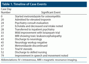
Neurologic workup of electroencephalography, lumbar puncture, infectious PCR (polymerase chain reaction) panel, multiple sclerosis profile, paraneoplastic panel, cerebrospinal fluid (CSF) cytology and flow cytometry, GAD65ab, and autoimmune encephalopathy panel were all negative except for elevated protein in the CSF. Brain magnetic resonance imaging (MRI) was pursued, which showed hyperintensity throughout the supratentorial white matter, sparing the gray matter and deep gray nuclei on MRI, with stable repeat imaging 17 days later. The workup and radiologic findings led to a diagnosis of metronidazole-induced toxic leukoencephalopathy with catatonia. He was taken off metronidazole and discharged to a skilled nursing unit, wherein he was noted to communicate in phrases, had improved stiffness, and was no longer posturing (Table 1).
Discussion
This case is unique for several reasons: (1) catatonia as a sentinel symptom of leukoencephalopathy, (2) catatonia and leukoencephalopathy as an adverse effect of metronidazole, and (3) catatonia not solely as a psychiatric presentation. Metronidazole has been known to cause leukoencephalopathy,1 though these patients typically present with dysarthria, ataxia, altered mental status, and, less commonly, seizures.2 With respect to psychiatric symptoms, metronidazole has been associated with psychosis due to its inhibition of bovine monoamine oxidase leading to increased levels of dopamine.3 Additionally, dopaminergic drugs such as cocaine or methadone have presented with leukoencephalopathy and catatonia.4,5 This case illustrates the copresentation of the known side effect of toxic leukoencephalopathy and previously undescribed catatonia with metronidazole toxicity. Metronidazole has been proposed to cause neuronal toxicity due to generated reactive oxygen species resulting in axonal edema, often reversible within days after discontinuing the offending agent.6 We recommend that patients presenting with catatonia should receive a thorough and complete medical workup. Not all cases of catatonia are solely caused by an underlying psychiatric condition. Workup should involve thorough clinical assessment, laboratory tests, and neuroimaging for rapid identification of the ongoing toxic process. Although removing the toxic drug should lead to improvement, treatment options such as benzodiazepines, N-methyl-D-aspartate receptor antagonists, or ECT may be warranted.
Published online: September 23, 2021.
Potential conflicts of interest: None.
Funding/support: None.
Previous presentation: This case was presented as an electronic poster (due to COVID-19) at the University of Kansas School of Medicine-Wichita Annual Research Forum; April 28, 2020; Wichita, Kansas.
Additional information: Information was de-identified to protect anonymity.
References (6)

- Roy U, Panwar A, Pandit A, et al. Clinical and neuroradiological spectrum of metronidazole induced encephalopathy: our experience and the review of literature. J Clin Diagn Res. 2016;10(6):OE01–OE09. PubMed CrossRef
- Sørensen CG, Karlsson WK, Amin FM, et al. Metronidazole-induced encephalopathy: a systematic review. J Neurol. 2020;267(1):1–13. PubMed CrossRef
- Befani O, Grippa E, Saso L, et al. Inhibition of monoamine oxidase by metronidazole. Inflamm Res. 2001;50(suppl 2):S136–S137. PubMed
- van Esch AMJ, Fest A, Hoffland BS, et al. Toxic leukoencephalopathy presenting as lethal catatonia. J Addict Med. 2019;13(3):241–244. PubMed CrossRef
- Anbarasan D, Campion P, Howard J. Drug-induced leukoencephalopathy presenting as catatonia. Gen Hosp Psychiatry. 2011;33(1):85.e1–85.e3. PubMed CrossRef
- Li L, Tang X, Li W, et al. A case of methylprednisolone treatment for metronidazole-induced encephalopathy. BMC Neurol. 2019;19(1):49. PubMed CrossRef
Enjoy this premium PDF as part of your membership benefits!


