Prim Care Companion CNS Disord 2022;24(2):21cr02969
To cite: Spiegel DR, McLean A, Swiacki NL, et al. Neuropsychiatric systemic lupus erythematosus presenting as mania with psychosis treated successfully with intravenous immunoglobulin: a diagnosis of associations and exclusions. Prim Care Companion CNS Disord. 2022;24(2):21cr02969.
To share: https://doi.org/10.4088/PCC.21cr02969
© Copyright 2022 Physicians Postgraduate Press, Inc.
aDepartment of Psychiatry and Behavioral Sciences, Eastern Virginia Medical School, Norfolk, Virginia
*Corresponding author: David R. Spiegel, MD, Department of Psychiatry and Behavioral Sciences, Eastern Virginia Medical School, 825 Fairfax Ave, Norfolk, VA 23507 ([email protected]).
The American College of Rheumatology (ACR) nomenclature provides case definitions, evaluations, and assessments for 19 neuropsychiatric/5 psychiatric syndromes of neuropsychiatric systemic lupus erythematosus (NPSLE).1,2 We present the case of a patient with new-onset mania, with unreported elevated anti-double stranded (ds) DNA and SLE.
CASE REPORT
Our patient was a 22-year-old Black woman who presented under an involuntary admission to our psychiatry unit with a 2-week history of grossly “unusual behavior.” Her mood was elated with excessive energy, auditory hallucinations, and persecutory/grandiose delusions. She scored 37 on the Young Mania Rating Scale (YMRS).3 On admission, urine drug screen was remarkable only for cannabis, and her blood alcohol screen was negative. The patient had no personal or family history of psychiatric illness. Upon admission, olanzapine was started for new-onset mania with psychotic features.
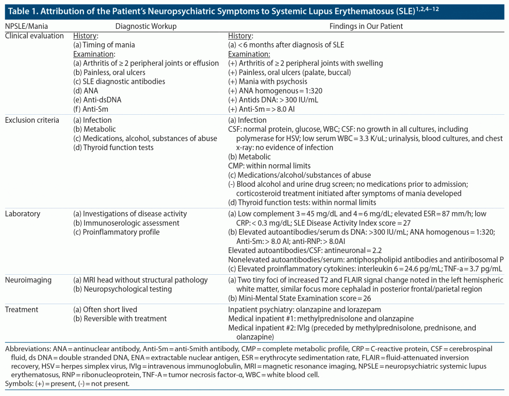
It was revealed that 5 months earlier when the patient was hospitalized for gastric ulcer, anti-dsDNA of 288I U/mL was listed without further evaluation. Thus, the patient was transferred to the medical service, and the consult service was consulted. Table 1 provides the results of the patient’s evaluation.
The patient was started on prednisone 60 mg/d, hydroxychloroquine 200 mg/d, and azathioprine 50 mg/d but refused olanzapine. After 3 days of treatment, with no change in YMRS score, methylprednisolone 500 mg twice/d was added; however, no change was observed 5 days later. Hence, both methylprednisolone and prednisone were tapered. Two days later, intravenous immunoglobulin (IVIg) (2 g/kg over 2 days) was initiated. Subsequently, 1 and 2 days after IVIg therapy was completed, her YMRS score decreased to 12 and 4, respectively. Four days after IVIg was completed, the patient was discharged with a YMRS score of 1. At 2-month follow-up, the patient remained adherent to immunosuppressants with no relapse of NPSLE. The diagnosis of SLE was based on our patient meeting the major criteria as shown in Table 1.1,2,4–12 Furthermore, please see Table 1 to review our rationale in attributing our patient’s mania to NPSLE.
DISCUSSION
Since there are no definitive serum or imaging biomarkers of NPSLE, attribution of NPSLE in our patient was based on the Systemic Lupus International Collaborating Clinics NPSLE inception cohort.4 Interestingly, NPSLE is estimated to occur before or around the time of the SLE diagnosis in 28%–40% of adults and in 63% within the first year after diagnosis.6 Our patient’s NPSLE occurred ~ 3 months after SLE diagnosis. Additionally, the stipulation of concurrent non‐SLE factor(s) could have been complicated had corticosteroids been initiated prior to the development of mania with psychotic features in our patient.13 Lastly, as mania with psychotic features is not a “common” NP event in normal population controls,5 we felt that our patient’s phenomenology surpassed these final criteria.
We would be remiss to not mention the consideration of a primary mood or psychotic disorder as a potential contributing factor/association. While the mean age at onset of bipolar disorder and SLE are similar,7,14 Black women are disproportionately afflicted with the latter.11 Notably, there is no significant difference in rates of bipolar disorder by race.15 Ultimately, our patient’s temporal proximity of mania to the diagnosis of SLE skewed our impression toward NPSLE.
Finally, there is no definitive treatment for NPSLE; although similar to other autoimmune encephalitides, we utilized a stepwise escalation of immunotherapy.16,17 While treatment response should not be used to confirm a diagnosis, our patient’s response to immunomodulation was supportive, albeit not conclusive, that her episode was due to NPSLE and not primary mania with psychotic features.
As we have shown, attribution of diffuse/neuropsychiatric events to SLE is a multifaceted process. While revisions and algorithms have been made to modify original criteria set by the ACR, diagnosis is based on a combination of psychiatric/neurologic evaluation, magnetic resonance imaging (MRI), and cerebrospinal fluid analysis.
NPSLE/Mania
In 1999, the ACR developed a standardized nomenclature system for the neuropsychiatric syndromes of NPSLE. Case definitions including diagnostic criteria, important exclusions, and methods of ascertainment were developed for 19 NPSLE syndromes, including mood disorders such as mania.1
Clinical Evaluation
Perhaps the most important piece of historical data about any neuropsychiatric symptom, including our patient’s mania, is its temporal relationship with respect to SLE clinical onset: highest in the 6 months prior to and in the first year following the diagnosis of SLE.2,4
According to the Systemic Lupus International Collaborating Clinics rule for the classification of SLE, our patient had at least 4 criteria, including at least 1 clinical criterion and 1 immunologic criterion.5
Exclusion Criteria
Since there are no definitive serum or imaging biomarkers of NPSLE, attributing neuropsychiatric events to SLE requires other potential causes (exclusions) and contributing factors (associations) to be evaluated and potentially ruled out. Our patient’s workup resulted in an exclusion of other causes of mood or psychotic disorder: (1) infections (eg, central nervous system), (2) hypertensive, (3) metabolic causes, and (4) medication/substance‐ or drug‐induced mood/psychotic disorder.
Other associations could include primary mood or psychotic disorder unrelated to SLE (eg, bipolar disorder with psychotic features, schizoaffective disorder) and, less likely, a psychologically mediated reaction to SLE (brief psychotic disorder).1,6,7
Laboratory
There are no specific and sensitive serum or imaging biomarkers of NSLE. Our patient’s antiribosomal P was negative. Antiribosomal P is inconsistently but associated with psychosis attributed to SLE.6 Our patient had highly elevated antidsDNA.
While a review of biomarkers is beyond the scope of this report, one promising biomarker identified for NPSLE is antidsDNA/NMDA-NR2. The latter are widely distributed in the brain, mostly in the hippocampus and amygdala, structures that control cognitive functions and emotional reactions, respectively.8
In NPSLE, anti-NMDAR/NR2 antibodies represent 40% of antidsDNA.9 While our patient’s level of antidsDNA was highly elevated, this does not prove that our patient’s antidsDNA cross-reacted with NR2 receptors in the central nervous system.
Our patient did have an elevated IL-6 level. Of the many cytokines proven to be present in cerebrospinal fluid, IL-6 has the strongest positive correlation with NPSLE.10
Neuroimaging
Our patient did have white matter hyperintensities, one of the common, albeit nonspecific, findings on MRI of her head. White matter hyperintensities have been shown to involve preferentially the frontal and parietal lobes, consistent with an anterior to posterior gradient.11
Our patient did not have cortical atrophy, the other most common MRI finding in NPSLE. Two factors that are associated with cortical atrophy are longer disease duration and cognitive dysfunction; neither were present in our patient.11
Treatment
The one associative factor in our patient that could not be conclusively ruled out (cross-sectionally) by our evaluation was the possibility of a primary mood or even psychotic disorder. Treatment should not be used to establish a diagnosis, although our patient was treated without psychotropics. While there is evidence of immune system overactivation in several psychiatric conditions, no evidence of immunotherapy to treat primary psychiatric disorders was found.12
CONCLUSION
Mania in our patient with conclusive SLE developed within the requisite time period to be consistent with NPSLE. Exclusionary and associative causes were considered and determined to be unlikely in the pathogenesis of mania. Antiribosomal P, laboratory data, and neuroimaging were sensitive, albeit not specific, to NPSLE. Thus, while it is acknowledged that current diagnostic assessment methods need improvement, we feel our patient did indeed have mania due to SLE.
Published online: March 15, 2022.
Potential conflict of interests: Dr Spiegel is in the speakers’ bureaus for Allergen, Alkermes, Otsuka, and Intra-Cellular Therapies, but has no conflicts of interest related to the subject of this report. Drs McLean, Swiacki, and Moran report no conflicts of interest.
Funding/support: None.
Patient consent: Consent was verbally received from the patient to publish the case report, and information has been de-identified to protect anonymity.
References (17)

- The American College of Rheumatology nomenclature and case definitions for neuropsychiatric lupus syndromes. Arthritis Rheum. 1999;42(4):599–608. PubMed CrossRef
- Meszaros ZS, Perl A, Faraone SV. Psychiatric symptoms in systemic lupus erythematosus: a systematic review. J Clin Psychiatry. 2012;73(7):993–1001. PubMed CrossRef
- Young RC, Biggs JT, Ziegler VE, et al. A rating scale for mania: reliability, validity and sensitivity. Br J Psychiatry. 1978;133(5):429–435. PubMed CrossRef
- Hanly JG, Li Q, Su L, et al. Psychosis in systemic Lupus Erythematosus: results from an International Inception Cohort Study. Arthritis Rheumatol. 2019;71(2):281–289. PubMed CrossRef
- Ainiala H, Hietaharju A, Loukkola J, et al. Validity of the new American College of Rheumatology criteria for neuropsychiatric lupus syndromes: a population-based evaluation. Arthritis Rheum. 2001;45(5):419–423. PubMed CrossRef
- Popescu A, Kao AH. Neuropsychiatric systemic lupus erythematosus. Curr Neuropharmacol. 2011;9(3):449–457. PubMed CrossRef
- Joo YB, Park SY, Won S, et al. Differences in clinical features and mortality between childhood-onset and adult-onset systemic Lupus Erythematosus: a prospective single-center study. J Rheumatol. 2016;43(8):1490–1497. PubMed CrossRef
- Arinuma Y, Yamaoka K. Developmental process in diffuse psychological/neuropsychiatric manifestations of neuropsychiatric systemic lupus erythematosus. Immunol Med. 2021;44(1):16–22. PubMed CrossRef
- Nikolopoulos D, Fanouriakis A, Boumpas DT. Update on the pathogenesis of central nervous system lupus. Curr Opin Rheumatol. 2019;31(6):669–677. PubMed CrossRef
- Cozzani E, Drosera M, Gasparini G, et al. Serology of Lupus Erythematosus: correlation between immunopathological features and clinical aspects. Autoimmune Dis. 2014;2014:321359. PubMed CrossRef
- Lim SS, Drenkard C. Epidemiology of lupus: an update. Curr Opin Rheumatol. 2015;27(5):427–432. PubMed
- Forrester A, Latorre S, O’Dea PK, et al. Anti-NMDAR Encephalitis: a multidisciplinary approach to identification of the disorder and management of psychiatric symptoms. Psychosomatics. 2020;61(5):456–466. PubMed CrossRef
- Bhangle SD, Kramer N, Rosenstein ED. Corticosteroid-induced neuropsychiatric disorders: review and contrast with neuropsychiatric lupus. Rheumatol Int. 2013;33(8):1923–1932. PubMed CrossRef
- Bellivier F, Golmard JL, Rietschel M, et al. Age at onset in bipolar I affective disorder: further evidence for three subgroups. Am J Psychiatry. 2003;160(5):999–1001. PubMed CrossRef
- Akinhanmi MO, Biernacka JM, Strakowski SM, et al. Racial disparities in bipolar disorder treatment and research: a call to action. Bipolar Disord. 2018;20(6):506–514. PubMed CrossRef
- Milstone AM, Meyers K, Elia J. Treatment of acute neuropsychiatric lupus with intravenous immunoglobulin (IVIG): a case report and review of the literature. Clin Rheumatol. 2005;24(4):394–397. PubMed CrossRef
- Brogna C, Manna R, Contaldo I, et al. Intravenous immunoglobulin for pediatric neuropsychiatric lupus triggered by Epstein-Barr Virus cerebral infection. Isr Med Assoc J. 2016;18(12):763–766. PubMed
Please sign in or purchase this PDF for $40.
Save
Cite


